Updated February 25, 2022
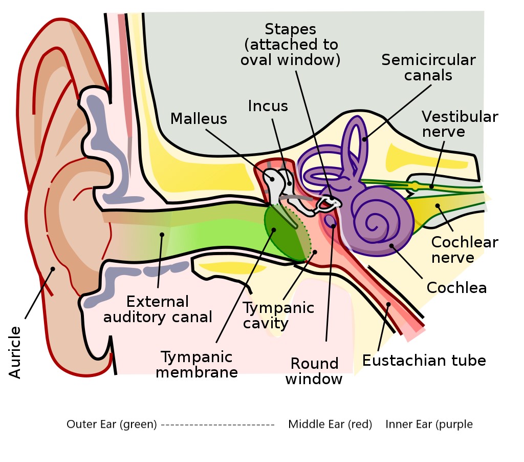
The human ear is responsible for our auditory sense (hearing) and our balance. It consists of three anatomical sections – the inner ear, middle ear, and outer ear.
Outer Ear
The outer ear consists of two parts:
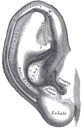
- The visible part of the ear, called the pinna. The pinna includes the auricle, the upper portion of the visible ear, and the ear lobe, the lower portion of the visible ear. (Though sometimes “auricle” and “pinna” are used interchangeably). The auricle contains elastic cartilage, called the auricular cartilage, which lacks blood vessels and nerve endings. It is a permanent cartilage that does not regrow or replace itself, so damage is almost always permanent. It is structured in such as way to aid in sound wave collection.
- The ear canal, which is a tunnel made of skin and bone called the external acoustic meatus that enters the skull. Glands in the outer third of the ear canal produce cerumen, or earwax. It is a moisturizing, cleansing, antibacterial agent that helps keep the path clear for sound waves and prevent infection. Under normal circumstances, the wax naturally leaves the ear due to the actions of the muscles of the jaw and mouth (including the levator veli palatini).
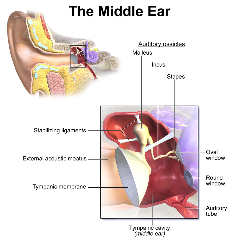
Middle Ear
The middle ear is a pressurized, membrane-lined, air-filled cavity separating the inner ear from the external environment and is known as the tympanic cavity. The pressure is maintained by the eustachian (or pharyngotympanic) tube. The tympanic cavity contains the eardrum and ear bones.
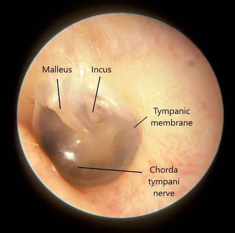
- The eardrum separates the outer and middle ear. It is a tympanic membrane measuring roughly one-tenth of a millimeter thick and a third of an inch in diameter, and is held in place by a ring of cartilage. The membrane consists of three layers of tissue: outer cutaneous, middle fibrous, and inner mucous membrane.
- Behind the membrane are ossicles, three tiny bones that create the ossicular chain. These are the tiniest bones in the human body and delicate enough to transmit vibrations from the tympanic membrane to the inner ear.
The ear bones
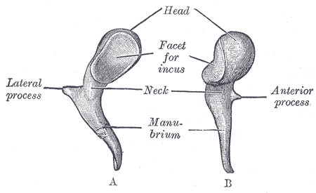
- The malleus bone (or hammer) is approximately 8 mm long in adult humans and is attached to the tensor tympani muscle, which allows regulation of loud noises to prevent damage to the eardrum. This process is called the acoustic reflex. The malleus bone has a lateral and anterior process of the manubrium (or handle), a head that connects to the incus bone via a facet, and a neck that connects the head and manubrium
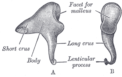
- The incus bone (or anvil) also has a facet where it contacts the head of the malleus. The incus consists of a short and long crus, with a curved lenticular process at the end of the long crus where it connects with the stapus bone. The center of the incus bone is referred to as the body.
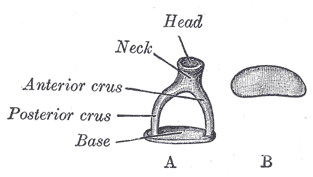
- The stapes (or stirrup) is the smallest bone at less than a third of a centimeter in length and is stabilized by the stapedius muscle. This bone consists of a head that makes contact with the lenticular process of the incus bone and a flat base that makes contact with the oval window. Connecting these structures is a neck: the inferior and superior (or anterior and posterior) crus.
Inner Ear
The inner ear is a fluid-filled structure containing the auditory and vestibular systems. The outer membrane of the inner ear is the oval window, or fenstra ovalis. The stapes contacts this membrane. Behind the oval window is the vestibule, from which the cochlea, semicircular canals, utricle, and saccule branch off.
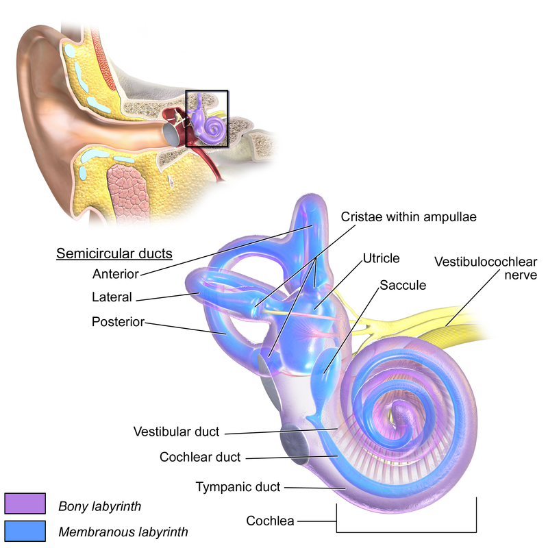
The cochlea
The spiral-shaped cochlea is comprised of three canals (scala) wrapped around the bony axis (the modiolus): the scala media (or cochlear duct), scala vestibule, and scala tympani.
- The so-called bony labyrinth includes the scalae vestibule and tympani and contains perilymph. This fluid is similar in composition to cerebrospinal fluid. These two canals connect at the apex (opening) of the cochlea at the helicotrema.
- The so-called membranous labyrinth is between the the two canals of the bony labyrinth. It comprises the triangular-shaped scala media and is filled with endolymph. This fluid has ion concentrations similar to intracellular fluid.
Inside the cochlea, on the basilar membrane between the scala media and scala tympani, is the organ of Corti, which contains the inner and outer hair cells. These hair cells are attached to nerve endings that convey sound information to the brain via the vestibulocochlear nerve (cochlear nerve VIII, also known as the auditory nerve).
The round window is a membrane located in the lower portion of the cochlea. It acts as a pressure release when sound waves push the oval window above it.
The vestibular system
Branching off the vestibule with membranous labyrinth, and containing endolymph, are the otolith organs. In humans these are the utricle and saccule. Otoliths are calcium carbonate structures that respond to movement and allow you to respond to gravity and acceleration.
Also connected via the membranous labyrinth are three semicircular canals lined with cilia and filled with endolymph. This is where nerve endings enter the vestibular system (cranial nerve VIII). Each semicircular canal provides a different directional sensor: horizontal, posterior, and superior.
Disclaimer: This page is for informational and learning purposes only. It is not meant to diagnose or treat any medical condition and should not be used in place of speaking with a medical doctor or seeking treatment.
Aliconia Publishing, LLC and the author make any and all attempts to ensure the accuracy of the presented facts. If you find an issue with any information on these pages, please use the Contact page to alert us. The content is subject to change based on new information or to be updated with additional facts. The date of last change is stated under the main header.
Discover more from Just Facts
Subscribe to get the latest posts sent to your email.