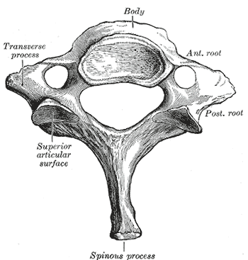
The cervical spine consists of the first seven vertebrae of the spinal column (denoted as C1-C7), starting at the base of the skull. This part of the spine provides support for the head and creates the neck, which provides the ability to rotate the head at the top of the spine.
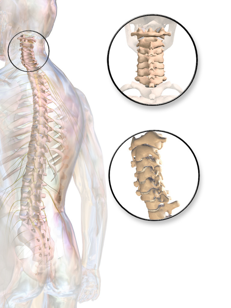
The vertebrae of the spine act as a protective bony sheath for the spinal cord, as well as nerves and blood vessels that branch off from the main vessels to and from the head. The cervical vertebrae are smaller than the vertebrae elsewhere in the spinal column, but they have the same general construction of a vertebral disc between each, supported by ligaments, tendons, and muscles.
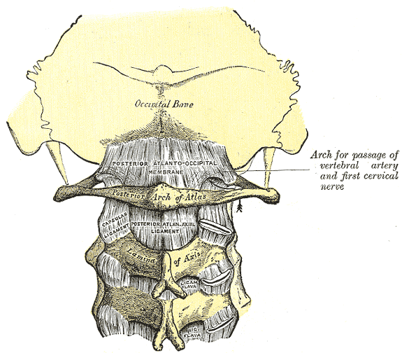
Atlas and axis (C1 and C2)
An exception to the standard vertebral structure is C1 and C2. The first two vertebrae at the top of the spinal column are unique in that they fit into one another. C1, called the atlas, is a ring that fits over the odontoid process of C2, called the axis. However, this interlocking allows rotation of the head.
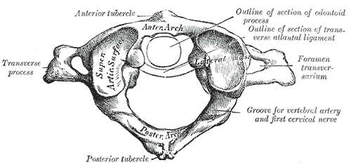
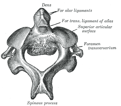
The connection between the spine and cranium is called the craniovertebral junction. The joint made by C1-2 and the occipital bone is the atlanto-occipital joint.
As anatomical markers, C1 is considered to be at the level of the hard palate/base of the nose and C2 at the teeth when the mouth is closed.
C3-C7
The rest of the vertebrae in the cervical spine have the same bone structure as the other vertebrae. An exception is the relatively long spinous process of C7, the vertebra prominens.
Most neck movements are controlled by C5-C7. Diseases or injury to the spine can cause fusion of the vertebrae (i.e., stenosis), eliminating the capacity to move the head and neck.
As anatomical markers, C3 is at the level of the hyoid bone/mandible, C4 the bifurcation of the common carotid artery, C4-5 thyroid cartilage, C6 where the esophagus, larynx, and trachea separate, C6-7 the cricoid cartilage. The carotid pulse is taken at the transverse process of C6.
Cervical nerves
Eight cervical nerves (also denoted as C1-C8) of the peripheral nervous system branch off from the spinal cord in the cervical spine. Each controls a different region of the upper body, including movement, sensory innervations, and body functions, though the exact control each exerts and how their functions overlap can vary. In general:
- Nerves C1 and C2 control the head and neck, as well as feeling on the face and scalp.
- Nerve C3 controls the diaphragm, and thus the muscles involved in breathing. It may also overlap with C1-2 more than with C4.
- Nerve C4 controls the upper body muscles of the shoulders and chest, including the ribs and diaphragm for expansion and contraction during breathing.
- Nerve C5 controls the upper body muscles of the shoulders and arms, including the deltoids and biceps.
- Nerve C6 controls the wrist extensors and some innervation to the biceps, as well as feeling in the thumb and hand.
- Nerve C7 controls the triceps and feelings down to the middle finger.
- Nerve C8 controls the hands and feeling in the forearm and pinky finger.
Muscles of the Cervical Spine
There are three sets of muscles that act on the cervical spine – anterior neck muscles, posterior neck muscles, and lateral neck muscles.
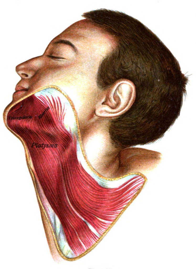
Anterior neck muscles
In addition to head movement, these muscles also play a role in swallowing, speech, and breathing.
- Infrahyoids (including thyrohyoid, sternohyoid, sternothyroid, and omohyoid) – control the larynx
- Playtsma – jaw and mouth movement
- Scalene – stabilization of cervical spine
- Sternocleidomastoid – neck extension and head movement, runs from behind the ear to the collarbone
- Subclavian – stabilizes collarbone
- Suprahyoids (including mylohyloid, stylohyloid, digastric, and geniohyoid) – move the hyoid to aid in chewing, speaking, swallowing
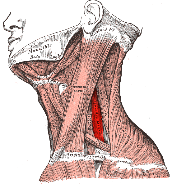
Posterior neck muscles
These muscles are at the back of the neck, extending towards the shoulders on both sides to assist in movement and extension of the head and neck.
- Splenius capitis
- Splenius cervicus
- Suboccipital muscles (including Rectus capitis posterior and Obliquus capitis)
- Transversospinalis muscles – also provide spinal stability
Lateral neck muscles
Also referred to as prevertebral muscles, these muscles are found on either side of the neck. They are responsible for rotation of the head.
- Longus capitalis
- Longus colli
- Rectus capitis (anterior and lateralis)
Disclaimer: This page is for informational and learning purposes only. It is not meant to diagnose or treat any medical condition and should not be used in place of speaking with a medical doctor or seeking treatment.
Aliconia Publishing, LLC and the author make any and all attempts to ensure the accuracy of the presented facts. If you find an issue with any information on these pages, please use the Contact page to alert us. The content is subject to change based on new information or to be updated with additional facts. The date of last change is stated under the main header.