Updated July 5, 2024
The Heart Muscle
- Myocytes – The individual cells of the heart.
- Myocardial septum – Tissue separating the heart chambers.
- Ventricles – The larger two chambers in the lower portion of the heart, known as the right and left ventricle. The ventricles are separated by the interventricular septum; valves separate the ventricles from the pulmonary artery (right) and aorta (left).
- Atria – The upper two chambers of the heart, known as the right atrium and left atrium. The atria are separated from the ventricles by valves, and from each other by the atrial septum, which joins with the interventricular septum immediately after birth by the foramen ovale. During fetal development, blood flows through a hole in the atrial septum due to a lack of blood oxygenation by the lungs prior to birth.
- Pericardial sac – The fibrous membrane surrounding the myocardium.
Heart Valves
- Valve – A one-way flap preventing the backflow of blood. There are four valves in the heart.
- Aortic valve – Separates the left ventricle and the aorta.
- Pulmonary valve – A semilunar (describing the shape: half moon) valve separating the pulmonary artery and the right ventricle.
- Tricuspid valve – Separates the right atrium and right ventricle.
- Mitral valve – A bicuspid valve between the left atrium and left ventricle.
- Cuspids – The tissue flaps that make a valve. Bi- and tri-cuspid refer to the number of flaps (two and three, respectively).
- Chordae – Tendon that holds the valve flaps taunt, preventing backflow when the heart contracts.
Blood Vessels
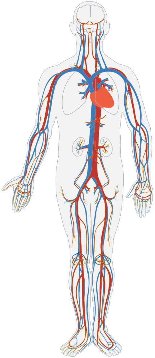
- Artery –Carries blood, mostly oxygenated, from the heart to the tissues. The pulmonary artery is the only one to carry deoxygenated blood, which is from the heart to the lungs. (mnemonic: A for Away)
- Vein – Carries blood, mostly deoxygenated, from the tissues to the heart. The pulmonary vein is the only one to carry oxygenated blood, which is from the lungs to the heart.
- Aorta – The largest artery; a muscular blood vessel stemming from the left ventricle. The aorta forms an arch and runs down to the abdomen. Other arteries branch off from the aorta to take blood to the head, the myocardium, the trunk, and the extremities.
- Coronary arteries – The arteries serving the outer myocardium.
- Vena cava – The veins that empty directly into the right atrium, known as the superior (upper, or anterior) and inferior (lower, or posterior) vena cava.
Words Relating to Heart Function
- Contraction – Also known as systole, the mechanical function that pumps blood out of the heart. Caused by involuntary electrical impulses in the specialized cells of the heart known as modified myocytes.
- Atrial systole – Contraction of both atria at the same time, pushing blood into the ventricles. The atria begin to relax as the ventricles contract.
- Ventricular systole – Contraction of the ventricles, pushing blood into the lungs and aorta.
- Diastole – Relaxation of the heart, allowing blood to flow into the atria.
- Node – Component of the electrical conductance system in the cardiac tissue.
- Sinoatrial (SA) node – The natural pacemaker of the heart, initiates systole in the right atrium.
- Atrioventricular (AV) node – In the atrial septum, receives a signal from the SA node.
- Purkinje fibers and the bundle of His – Other electrical conduction fibers that stimulate contraction of the ventricles.
Defects in Heart Function
- Arrhythmia – An irregular heart rhythm.
- Fibrillation – A type of arrhythmia caused by problems in the cardiac nodes that affect contraction.
- Atrial fibrillation – Results in inadequate emptying of the atria.
- Ventricular fibrillation – Inadequate ventricle contraction; a cause of sudden death.
- Myocardial infarction – Known as a heart attack; lack of blood flow through the coronary arteries resulting in myocardium damage (infarct) due to a lack of oxygenation.
- Cardiac tamponade – The pericardial sac fills with fluid, resulting in constriction of heart function.
Disclaimer: This page is for informational and learning purposes only. It is not meant to diagnose or treat any medical condition and should not be used in place of speaking with a medical doctor or seeking treatment.
Aliconia Publishing, LLC and the author make any and all attempts to ensure the accuracy of the presented facts. If you find an issue with any information on these pages, please use the Contact page to alert us. The content is subject to change based on new information or to be updated with additional facts. The date of last change is stated under the main header.
